What it isCommon
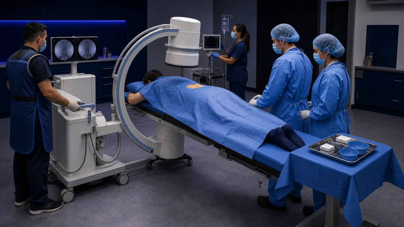
A contrast-enhanced X-ray examination
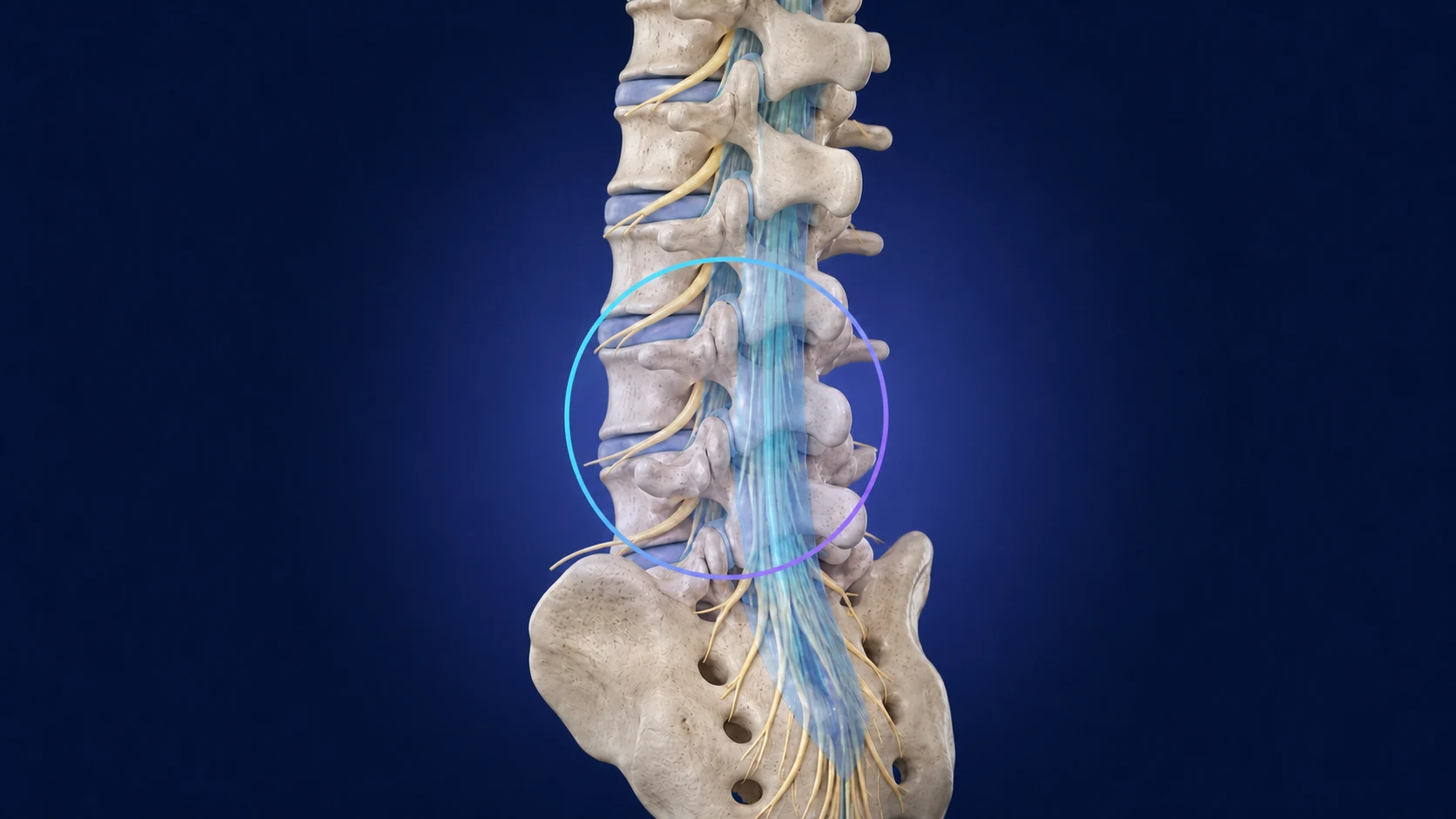
A qualified clinician places a spinal needle into the subarachnoid space and administers approved intrathecal iodinated contrast. Fluoroscopy shows the contrast within the subarachnoid space as it outlines the nerve roots, thecal sac, and related structures.